Where Compassion Inspires Progress

A karyotype examines the number and structure of chromosomes – which are the packages for your DNA. There should be 46 chromosomes present in each cell, 23 from mom and 23 from dad. A karyotype was the first test offered by the diagnostic laboratory at GGC in 1974 and is still a common test today to diagnose conditions such as Down syndrome.
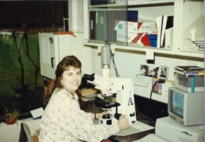
Bonne’ Lethco joined GGC as a technologist in the cytogenetics laboratory in 1989 and has spent the past 35 years preparing samples and analyzing chromosomes for thousands of patients.
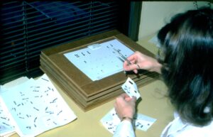
When Bonne’ started working at GGC, preparing the samples was a very long and a very manual multistep process. She used a microscope to count each chromosome and took pictures with a camera mounted to the microscope (with actual camera film!). Then the film was developed and the images were enlarged in the lab’s dark room. Finally, Bonne’ physically cut out each chromosome in the picture and pasted each one in order on paper to make a karyotype for analysis. She completed the initial analysis before sending it to the lab director who completed the final analysis and signed out the report. You can only imagine how long of a process this was!
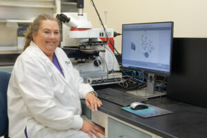
In 2016, the laboratory adopted a new technology, called CytoVision that reduced some of the hands-on manual steps and increased efficiency. CytoVision decreases the turnaround time and improves the resolution of chromosome images for better detection of abnormalities.
Bonne’ can now count each chromosome with a click of a mouse and digitally cut out each chromosome (think PhotoShop). She ensures the computer does the karyotype correctly (yes, even computers make mistakes), and she still does the initial analysis before the director reviews it.
While some of the processes for chromosome analysis has remained the same over the years (culturing cells, staining slides, conducting the initial analysis); the biggest and best change Bonne’ has seen with the addition of new technology is the decrease in turnaround time. When the cytogenetics lab adopted CytoVision, patients and families could receive a diagnosis in days rather than weeks.
Bonne’ Gives Greater Care by helping provide a diagnosis. She takes pride in knowing that having the correct diagnosis can lead to more resources or treatments for the patient. And although how we do the test has certainly changed over the years, our most important work is constant: our commitment to helping find answers for patients and their families.

©2024 Greenwood Genetic Center. All Rights Reserved. Privacy Policy. Notice of Nondiscrimination. Good Faith Estimate. Code of Conduct. Sitemap.